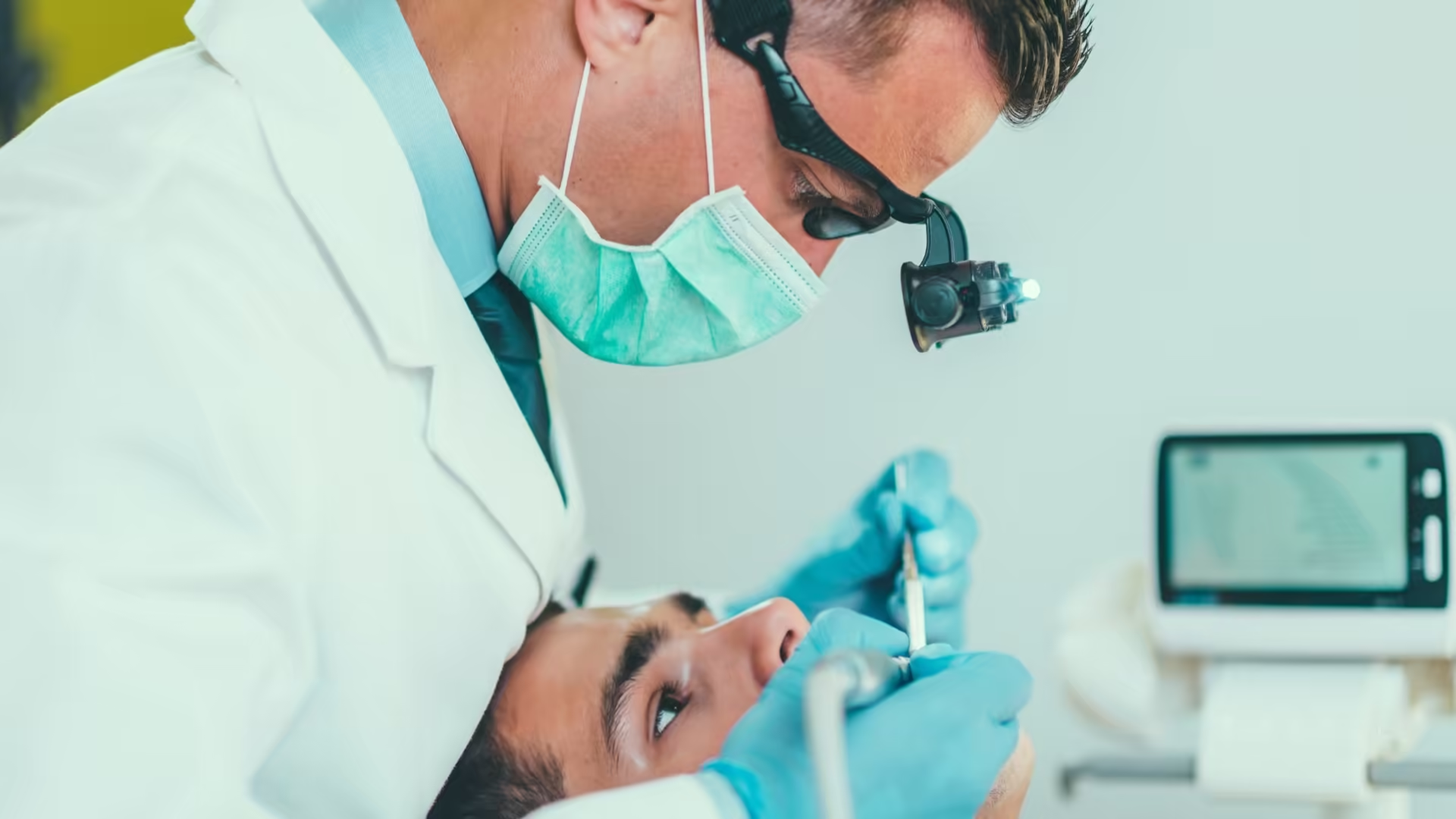




In brief: Gum disease is diagnosed by measuring the pockets around each tooth with a fine probe, checking for bleeding, and looking at bone levels on X-rays. Together these show if you have healthy gums, gingivitis or periodontitis, and if periodontitis, how advanced it is and how fast it is likely to progress. That diagnosis decides the treatment.
Why a gum diagnosis matters
Gum disease rarely hurts in its early stages, so it often goes unnoticed until it is advanced. Bleeding when brushing may be the only early sign, and many people ignore it. By the time teeth feel loose, a large share of the supporting bone may already be lost.
A proper periodontal examination finds the problem while it is still easier to treat. It is also a step before other dental work: crowns, implants, veneer work and orthodontic treatment all depend on healthy gums and bone, and active gum disease is usually treated first.
What the examination involves
Probing depths
The dentist slides a thin, blunt probe with millimetre markings gently into the space between gum and tooth, at several points around every tooth. In healthy gums this space is shallow, usually 1 to 3 mm. Readings of 4 mm or more suggest a pocket has formed, and deeper readings generally mean more advanced disease. The probing is mildly uncomfortable at most.
Bleeding on probing
Healthy gums rarely bleed when probed. Bleeding shows active inflammation at that site. Sites that keep bleeding over time are more likely to lose further attachment.
Attachment loss and recession
Pocket depth alone can mislead, because swollen gums make pockets look deeper and receding gums make them look shallower. The dentist also measures clinical attachment loss: how far the gum attachment has moved down the root from where it should be.
Mobility, furcations and plaque
Each tooth is checked for looseness. On back teeth with more than one root, the dentist checks the furcation, the area where the roots divide, for bone loss. Plaque and tartar levels are recorded because they show where cleaning is being missed.
X-rays and CBCT
Dental X-rays show the height of the bone around each tooth and the pattern of bone loss, whether even across several teeth or as deeper defects beside individual roots. Where more detail is needed, for example before regenerative surgery or implants, a CBCT scan gives a three-dimensional view of the bone. Dazzle has CBCT 3D imaging in-house.
All findings are written onto a periodontal chart. This becomes the baseline for judging whether treatment has worked.
Staging and grading in plain terms
Periodontitis is commonly classified in two ways under the current international system.
- Stage (I to IV) describes how severe and complex the disease is now, based on attachment loss, bone loss, teeth already lost to gum disease and how difficult treatment will be. Stage I is early; stage IV involves extensive damage and often affects chewing and tooth position.
- Grade (A to C) describes how fast the disease is progressing or is likely to progress. Smoking and poorly controlled diabetes push the grade higher.
A diagnosis such as "stage III, grade B periodontitis" tells the dentist both how much damage exists and how aggressively it needs to be managed.
What the results mean for you
- Healthy gums: shallow readings, little or no bleeding, normal bone levels. Routine cleaning and check-ups continue.
- Gingivitis: bleeding and inflammation but no attachment or bone loss. It is reversible with a professional clean and better home care.
- Periodontitis: pockets, attachment loss and bone loss. Bone already lost does not regrow on its own, but further loss can usually be slowed or stopped with treatment and maintenance.
Next steps after diagnosis
For periodontitis, the first step is almost always non-surgical: scaling and root planing, plus help with brushing and cleaning between teeth. Risk factors such as smoking and diabetes are addressed at the same time.
A few weeks later, the gums are measured again. If deep pockets persist, options include further cleaning, gum surgery to access and clean the roots, or regenerative procedures to rebuild lost bone in suitable defects. Once the disease is stable, maintenance visits every three to four months are common.
A periodontal assessment is also part of planning cosmetic and restorative work. Before crown lengthening, for example, the dentist checks gum thickness and bone levels to decide how much tissue can be reshaped while leaving enough attachment to keep the gums healthy.
Bleeding gums or loose teeth? Book a Consult at our Bandra West or Kemps Corner clinic for a full gum assessment.